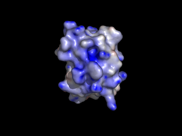
The structure will be processed by pdb2gmx, and you will be prompted to choose a force field:ġ: CHARMM36 all-atom force field (July 2017)įrom '/usr/local/gromacs/share/gromacs/top':Ģ: AMBER03 protein, nucleic AMBER94 (Duan et al., J. Gmx pdb2gmx -f 3HTB_clean.pdb -o 3HTB_oĬhoose the default water model when prompted.

Write the topology for the T4 lysozyme with pdb2gmx: There should now be a "charmm36-mar2019.ff" subdirectory in your working directory. Unarchive the force field tarball in your working directory:
#Pymol tutorial 10 and 11 download
Download the version of the conversion script that corresponds to your installed Python version (Python 2.x or 3.x). While there, download the latest CHARMM36 force field tarball and the "cgenff_charmm2gmx.py" conversion script, which we will use later. The force field we will be using in this tutorial is CHARMM36, obtained from the MacKerell lab website. At this point, preparing the protein topology is trivial. Then simply delete the JZ4 lines from 3HTB_clean.pdb. Since we will be preparing these two topologies separately, we must save the protein and JZ4 ligand into separate coordinate files. Prepare the ligand topology using external tools.Prepare the protein topology with pdb2gmx.Since this is not the case, we will prepare our system topology in two steps: rtp (residue topology) file for the force field. Topologies can only be assembled automatically if an entry for a building block is present in the. The problem we now face is that the JZ4 ligand is not a recognized entity in any of the force fields provided with GROMACS, so pdb2gmx will give a fatal error if you were try to pass this file through it. pdb file to check your work, you can download it here. We will instead focus on the ligand called "JZ4," which is 2-propylphenol.

For our intentions here, we do not need crystal water or other species, which are just crystallization co-solvents. Note that such a procedure is not universally appropriate (i.e., the case of a bound active site water molecule). Once you've had a look at the molecule, you are going to want to strip out the crystal waters, PO4, and BME. Once you have downloaded the structure, you can visualize it using a viewing program such as VMD, Chimera, PyMOL, etc. Go to the RCSB website and download the PDB text for the crystal structure.
#Pymol tutorial 10 and 11 code
For this tutorial, we will utilize T4 lysozyme L99A/M102Q (PDB code 3HTB). > Comply to PCI DSS 3.0 Requirement 10 and 11.We must download the protein structure file we will be working with. > Are you Audit-Ready for PCI DSS 3.0 Compliance? Download White paper > Achieve PCI DSS 3.0 Compliant Status with Out-of-the-box PCI DSS Reports > Meet PCI DSS 3.0 Compliance Requirements with EventLog Analyzer If you are not the named addressee you should not disseminate, distribute, copy, or alter this email. > This email and any files transmitted with it are confidential and intended solely for the addressee. > 2405 Wesbrook Mall, Fourth Floor | Vancouver, BC V6T 1Z3 | Main: > CDRD - The Centre for Drug Research and Development > How can I get PYMOL to use the CONECT table from a PDB file? My protein is glycosylated, and I'd like to properly and automatically display the glycosides including their linkage to the protein. > On Oct 6, 2014, at 6:35 PM, Markus Heller wrote: > From: David Hall Sent: Monday, Octo4:40 PM > But that doesn't work, shows no side chains. > This only shows me the glycoside, but NOT the side chain it's attached to. > Follow-up question: I want to show my glycosylated protein as cartoon, with the glycosides *and* the side chains they're attached to shown in sticks. Three-letter codes of the top of my head.

Galactose and sialic acid to that list I don't remember their The two places I have common monomer names for sugars.

NDG (because of an incorrect existing file from PDB), then add NDG to If this is your structure, I suggest you correct it. The PDB is replete with this mistake (about a third of N-linked sugarsĪre incorrectly named/modeled according to a paper by Thomas Lütteke). Usually an incorrectly modeled NAG (beta-N-acetyl-D-glucosamine), and Glycosylation, as far as I know, there are no NDG (alpha monomer of Select NlinkedGlyco, resn NAG+BMA+MAN+FUC or byres (polymer within 2.0Īlso, are you sure about NDG? If you are talking about N-linked


 0 kommentar(er)
